Tests
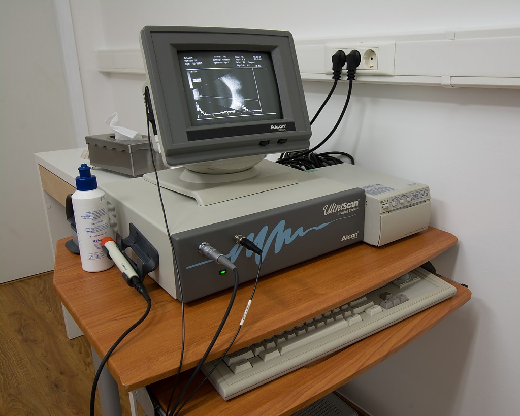
Ocular Ultrasound
Ultrasound or ocular ultrasonography is an imaging test that provides a direct, live view without the need for radiation and is easily practicable and reproducible. It can show the inside of the eye, even if an opacity of the media does not allow its visualization.
This test uses a very frequent sound, imperceptible to the human ear and absolutely innocuous. The ultrasound identifies any anatomical changes with a minimum size located in the vitreous cavity, the retina or the layer below (choroid), as well as in the soft tissues of the orbit (adipose tissue, oculomotor muscles) or the optic nerve. Any opacity in the anterior (cataract, hemorrhage) or posterior segment of the eye (vitreous cavity haemorrhage or vitreous turbidity) that avoids partial or total visualization of the retina, benefits from the use of this examination.
Preparation:
Usually no relevant preparation required.
